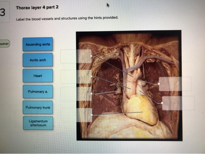
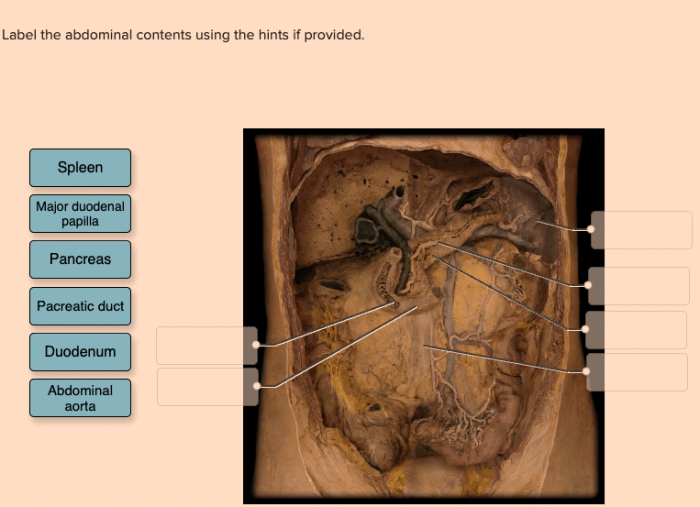
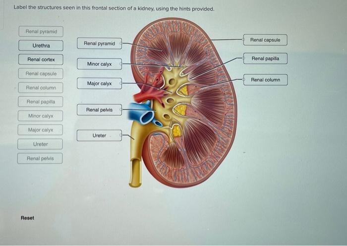
Label the blood vessels and structures using the hints provided – In the realm of medical imaging, the precise labeling of blood vessels and structures is paramount. This comprehensive guide will delve into the significance of labeling, explore various types of blood vessels and structures, and provide invaluable hints to ensure accurate identification.
By understanding the techniques and applications of labeling, medical professionals can enhance diagnosis, treatment, and research.
Labeling Blood Vessels and Structures

Labeling blood vessels and structures in medical imaging is crucial for accurate diagnosis and treatment. It allows medical professionals to visualize and identify specific anatomical features, facilitating better understanding of disease processes and guiding therapeutic interventions.
Blood vessels can be classified into arteries, veins, and capillaries. Arteries carry oxygenated blood away from the heart, while veins return deoxygenated blood to the heart. Capillaries are the smallest blood vessels, responsible for the exchange of nutrients and waste products between the blood and surrounding tissues.
In addition to blood vessels, other structures that may be labeled in medical imaging include bones, muscles, organs, and lymph nodes. These structures provide additional anatomical context and help in localizing and characterizing pathological findings.
Hints for Labeling Blood Vessels and Structures
Accurate labeling of blood vessels and structures relies on various hints. These hints can be anatomical landmarks, such as the location of specific bones or organs, or they can be based on the characteristics of the blood vessels themselves, such as their size, shape, and branching pattern.
For example, in computed tomography (CT) scans, the aorta, the largest artery in the body, can be identified by its large diameter and central location in the chest. In magnetic resonance imaging (MRI), veins can be distinguished from arteries based on their signal intensity, with veins typically appearing darker than arteries due to their slower blood flow.
Techniques for Labeling Blood Vessels and Structures
Several techniques can be used to label blood vessels and structures in medical imaging. These techniques include manual labeling, semi-automatic labeling, and fully automatic labeling.
Manual labeling involves manually tracing the contours of blood vessels and structures using a mouse or stylus. This method is time-consuming but provides precise and accurate results.
Semi-automatic labeling uses computer algorithms to assist in the labeling process. The user provides initial input, such as identifying a few key points, and the algorithm then generates a segmentation of the blood vessels or structures.
Fully automatic labeling algorithms can segment blood vessels and structures without any user input. These algorithms are still under development but have the potential to significantly reduce the time required for labeling.
Applications of Labeling Blood Vessels and Structures, Label the blood vessels and structures using the hints provided
Labeling blood vessels and structures has numerous applications in medical diagnosis and treatment.
- Disease diagnosis:Labeling blood vessels and structures can help diagnose a wide range of diseases, such as atherosclerosis, aneurysms, and tumors.
- Treatment planning:Accurate labeling is essential for planning surgical and other interventional procedures, such as stent placement and tumor resection.
- Medical research:Labeling blood vessels and structures enables researchers to study the anatomy and function of the vascular system and other organs.
Top FAQs: Label The Blood Vessels And Structures Using The Hints Provided
What is the significance of labeling blood vessels and structures in medical imaging?
Labeling blood vessels and structures allows for precise identification and visualization of anatomical features, aiding in diagnosis, treatment planning, and research.
How can hints help in labeling blood vessels and structures?
Hints provide clues based on anatomical relationships, shape, size, and location, assisting in accurate labeling and reducing errors.
What are the different techniques used for labeling blood vessels and structures?
Techniques include manual labeling, semi-automatic segmentation, and deep learning algorithms, each with its advantages and disadvantages.
How can labeling blood vessels and structures improve medical diagnosis and treatment?
Accurate labeling enables precise anatomical mapping, facilitating diagnosis, treatment planning, and monitoring of disease progression.


